Lens is made of glass.
Light microscope source of radiation.
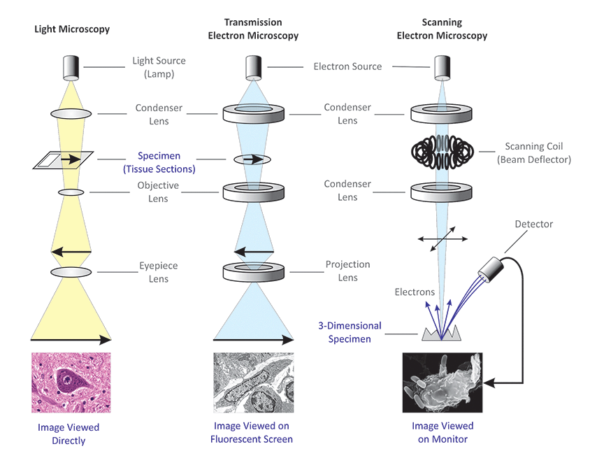
The sem is an instrument that produces a largely magnified image by using electrons instead of light to form an image.
Not affected by magnetic field.
This is confounded by the fact that most leds used in modern microscopes emit some uv light.
Since the light is in the visible range we can see images formed by a light microscope with naked eyes.
Light microscopes use light approx wavelength 400 700 nm electron microscopes use beams of electrons approx equivalent wavelength 1 nm.
There is risk of radiation leakage.
Light is focused with the help of glass lenses.
Specimen preparation takes usually takes few days.
Illuminating source is the light.
This has a wavelength of about 400 700 nm nanometer.
Illuminating source is the beam of electrons.
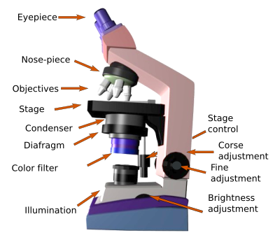
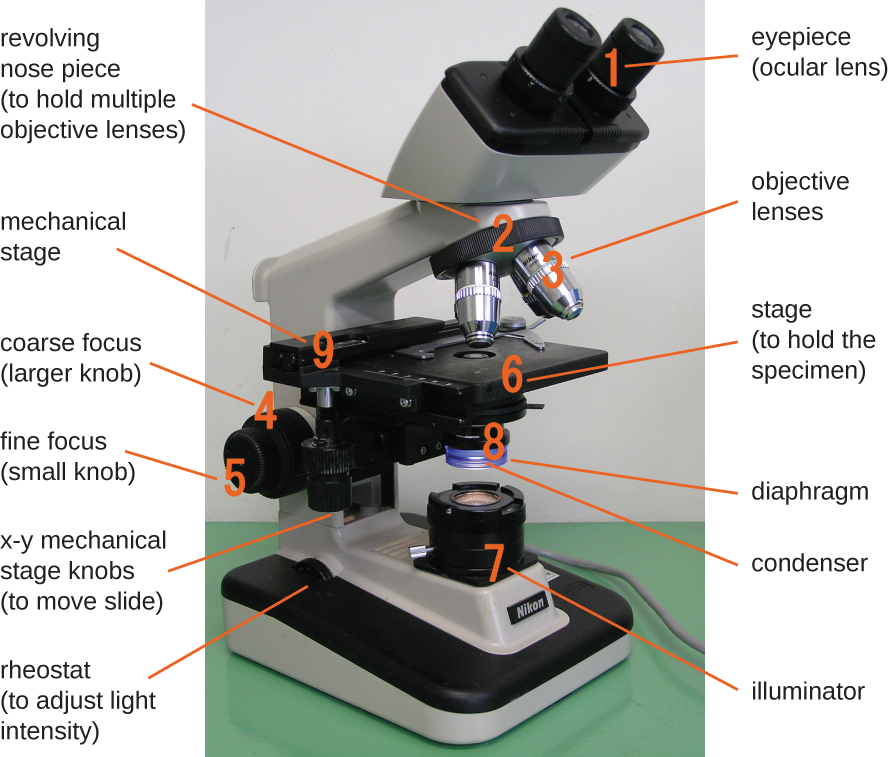
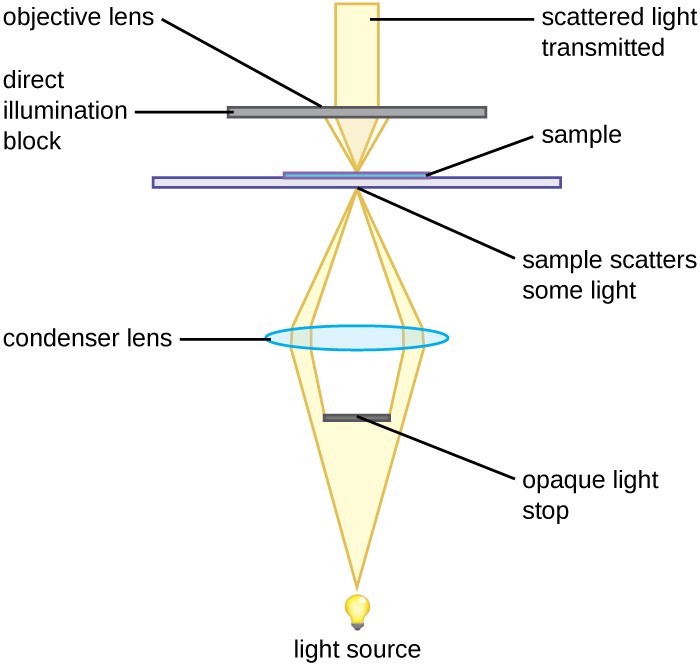
On most basic microscopes the diaphragm is located on top of the light source between the light bulb and the stage.
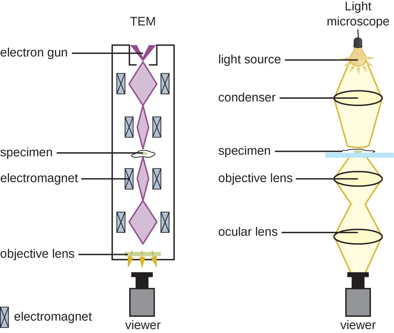
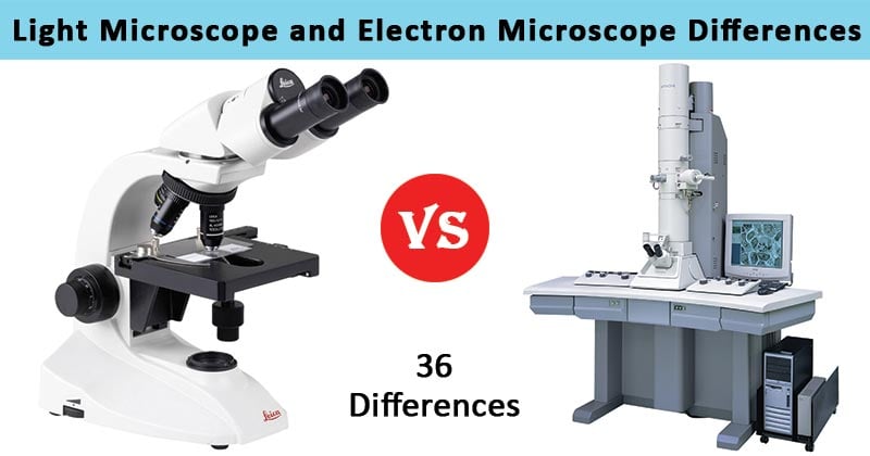
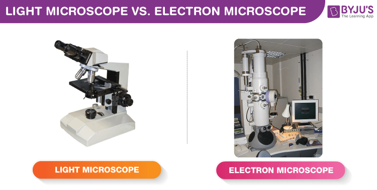
The most common types of microscopes are the light microscope and electron microscope.
A beam of electrons is produced at the top of the microscope by an electron gun.
X rays are produced in the electron microscope whenever the primary electron beam or back scattered electrons strike metal parts with sufficient energy to excite continuous and or characteristic x radiation.
The advanced light source als in berkeley california is home to xm 1 a full field soft x ray microscope operated by the center for x ray optics and dedicated to various applications in modern nanoscience such as nanomagnetic materials environmental and materials sciences and biology.
The electron beam follows a vertical path through the microscope which is held within a vacuum.
In general achromat lenses are the most basic whereas plan apochromats are often considered superior.
Specimen preparation takes usually few minutes to hours.
My inquiries revealed that plan apochromats have the highest levels of uv light transmission including to the eyes of the user.
Both light microscopes and electron microscopes use radiation to form detailed images of objects that a human eye cannot produce unaided.
Light via glass lenses beams of electrons can be focused using electromagnets due to negative charge on electrons.
The radiation source source of illumination is light wavelength 400 700 nm.
Control of image formation.
Source of radiation from an electron microscope.
Each of these microscopes possesses distinct features and is appropriate for different purposes.
1 nanometer 1 x 10 9 m.